Perisinusoidal space
| Perisinusoidal space | |
|---|---|
 | |
 Basic liver structure | |
| Details | |
| Location | Liver |
| Identifiers | |
| Latin | spatium perisinusoideum |
| TH | H3.04.05.0.00012 |
| Anatomical terms of microanatomy | |
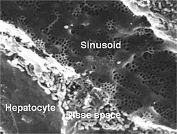
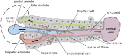
The perisinusoidal space (or space of Disse) is a space between a hepatocyte, and a sinusoid in the liver. It contains the blood plasma. Microvilli of hepatocytes extend into this space, allowing proteins and other plasma components from the sinusoids to be absorbed by the hepatocytes. Fenestration and discontinuity of the sinusoid endothelium facilitates this transport.[1] The perisinusoidal space also contains hepatic stellate cells (also known as Ito cells or lipocytes), which store vitamin A in characteristic lipid droplets.[2]
This space may be obliterated in liver disease, leading to decreased uptake by hepatocytes of nutrients and wastes such as bilirubin.
The Space of Disse is named for the German anatomist Joseph Disse (1852–1912).[3]
Pathophysiology
Fibrosis
Liver injury from a number of causes can activate the hepatic stellate cells into transdifferentiated and prolific myofibroblasts.[4] The myofibroblasts synthesize and secrete components of the extracellular matrix including collagen into the perisinusoidal space.[4] This in turn promotes the development of fibrosis, and continuing fibrosis is thought to be responsible for the development of cirrhosis, and liver cancer.[5]
References
- ^ Robbins, Stanley L.; Cotran, Ramzi S.; Kumar, Vinay; Collins, Tucker (1999). Robbins pathologic basis of disease. Philadelphia: Saunders. ISBN 0-7216-7335-X.
- ^ Kumar, Vinay; Abbas, Abul K.; Aster, Jon C.; Robbins, Stanley L.; Perkins, James A. (2018). Robbins basic pathology (Tenth ed.). Philadelphia, Pennsylvania: Elsevier. p. 637. ISBN 9780323353175.
- ^ Haubrich WS (2004). "Disse of the space of Disse". Gastroenterology. 127 (6): 1684. doi:10.1053/j.gastro.2004.10.021. PMID 15578505.
- ^ a b Tacke, F; Weiskirchen, R (February 2012). "Update on hepatic stellate cells: pathogenic role in liver fibrosis and novel isolation techniques". Expert Review of Gastroenterology & Hepatology. 6 (1): 67–80. doi:10.1586/egh.11.92. PMID 22149583.
- ^ Cheng, S; Zou, Y; Zhang, M (October 2023). "Single-cell RNA sequencing reveals the heterogeneity and intercellular communication of hepatic stellate cells and macrophages during liver fibrosis". MedComm. 4 (5): e378. doi:10.1002/mco2.378. PMC 10505372. PMID 37724132.
External links
- UIUC Histology Subject 1317
- Histology image: 22103loa – Histology Learning System at Boston University - "Ultrastructure of the Cell: hepatocytes and sinusoids, sinusoid and space of Disse"
- Histology image: 15204loa – Histology Learning System at Boston University - "Liver, Gall Bladder, and Pancreas: liver; sinusoids and Kupffer cells"
- Anatomy photo: digestive/mammal/liver5/liver7 - Comparative Organology at University of California, Davis - "Mammal, liver (EM, Low)"
