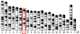
CD79A
| CD79A | |||||||||||||||||||||||||||||||||||||||||||||||||||
|---|---|---|---|---|---|---|---|---|---|---|---|---|---|---|---|---|---|---|---|---|---|---|---|---|---|---|---|---|---|---|---|---|---|---|---|---|---|---|---|---|---|---|---|---|---|---|---|---|---|---|---|
| |||||||||||||||||||||||||||||||||||||||||||||||||||
| Identifiers | |||||||||||||||||||||||||||||||||||||||||||||||||||
| Aliases | CD79A, IGA, MB-1, CD79a molecule, MB1, IGAlpha | ||||||||||||||||||||||||||||||||||||||||||||||||||
| External IDs | OMIM: 112205; MGI: 101774; HomoloGene: 31053; GeneCards: CD79A; OMA:CD79A - orthologs | ||||||||||||||||||||||||||||||||||||||||||||||||||
| |||||||||||||||||||||||||||||||||||||||||||||||||||
| |||||||||||||||||||||||||||||||||||||||||||||||||||
| |||||||||||||||||||||||||||||||||||||||||||||||||||
| |||||||||||||||||||||||||||||||||||||||||||||||||||
| |||||||||||||||||||||||||||||||||||||||||||||||||||
| Wikidata | |||||||||||||||||||||||||||||||||||||||||||||||||||
| |||||||||||||||||||||||||||||||||||||||||||||||||||
Cluster of differentiation CD79A also known as B-cell antigen receptor complex-associated protein alpha chain and MB-1 membrane glycoprotein, is a protein that in humans is encoded by the CD79A gene.[5]
The CD79a protein together with the related CD79b protein, forms a dimer associated with membrane-bound immunoglobulin in B-cells, thus forming the B-cell antigen receptor (BCR). This occurs in a similar manner to the association of CD3 with the T-cell receptor, and enables the cell to respond to the presence of antigens on its surface.[6]
It is associated with agammaglobulinemia-3.[7]
Gene
The mouse CD79A gene, then called mb-1, was cloned in the late 1980s,[8] followed by the discovery of human CD79A in the early 1990s.[9][10] It is a short gene, 4.3 kb in length, with 5 exons encoding for 2 splice variants resulting in 2 isoforms.[5]
CD79A is conserved and abundant among ray-finned fish (actinopterygii) but not in the evolutionarily more ancient chondrichthyes such as shark.[11] The occurrence of CD79A thus coincides with the evolution of B cell receptors with greater diversity generated by recombination of multiple V, D, and J elements in bony fish contrasting the single V, D and J elements found in shark.[12]
Structure
CD79a is a membrane protein with an extracellular immunoglobulin domain, a single span transmembrane region and a short cytoplasmic domain.[5] The cytoplasmic domain contains multiple phosphorylation sites including a conserved dual phosphotyrosine binding motif, termed immunotyrosine-based activation motif (ITAM).[13][14] The larger CD79a isoform contains an insert in position 88-127 of human CD79a resulting in a complete immunoglobulin domain, whereas the smaller isoform has only a truncated Ig-like domain.[5] CD79a has several cysteine residues, one of which forms covalent bonds with CD79b.[15]
Function
CD79a plays multiple and diverse roles in B cell development and function. The CD79a/b heterodimer associates non-covalently with the immunoglobulin heavy chain through its transmembrane region, thus forming the BCR along with the immunoglobulin light chain and the pre-BCR when associated with the surrogate light chain in developing B cells. Association of the CD79a/b heterodimer with the immunoglobulin heavy chain is required for surface expression of the BCR and BCR induced calcium flux and protein tyrosine phosphorylation.[16] Genetic deletion of the transmembrane exon of CD79A results in loss of CD79a protein and a complete block of B cell development at the pro to pre B cell transition.[17] Similarly, humans with homozygous splice variants in CD79A predicted to result in loss of the transmembrane region and a truncated or absent protein display agammaglobulinemia and no peripheral B cells.[7][18][19]
The CD79a ITAM tyrosines (human CD79a Tyr188 and Tyr199, mouse CD79a Tyr182 and Tyr193) phosphorylated in response to BCR crosslinking, are critical for binding of Src-homology 2 domain-containing kinases such as spleen tyrosine kinase (Syk) and signal transduction by CD79a.[20][21] In vivo, the CD79a ITAM tyrosines synergize with the CD79b ITAM tyrosines to mediate the transition from the pro to the pre B cell stage as suggested by the analysis of mice with targeted mutations of the CD79a and CD79b ITAM.[22][23] Loss of only one of the two functional CD79a/b ITAMs resulted in impaired B cell development but B cell functions such as the T cell independent type II response and BCR mediated calcium flux in the available B cells were intact. However, the presence of both the CD79a and CD79b ITAM tyrosines were required for normal T cell dependent antibody responses.[22][24] The CD79a cytoplasmic domain further contains a non-ITAM tyrosine distal of the CD79a ITAM (human CD79a Tyr210, mouse CD79a Tyr204) that can bind BLNK and Nck once phosphorylated,[25][26][27] and is critical for BCR mediated B cell proliferation and B1 cell development.[28] CD79a ITAM tyrosine phosphorylation and signaling is negatively regulated by serine and threonine residues in direct proximity of the ITAM (human CD79a Ser197, Ser203, Thr209; mouse CD79a Ser191, Ser197, Thr203),[29][30] and play a role in limiting formation of bone marrow plasma cells secreting IgG2a and IgG2b.[23]
Diagnostic relevance
The CD79a protein is present on the surface of B-cells throughout their life cycle, and is absent on all other healthy cells, making it a highly reliable marker for B-cells in immunohistochemistry. The protein remains present when B-cells transform into active plasma cells, and is also present in virtually all B-cell neoplasms, including B-cell lymphomas, plasmacytomas, and myelomas. It is also present in abnormal lymphocytes associated with some cases of Hodgkins disease. Because even on B-cell precursors, it can be used to stain a wider range of cells than can the alternative B-cell marker CD20, but the latter is more commonly retained on mature B-cell lymphomas, so that the two are often used together in immunohistochemistry panels.[6]
See also
References
- ^ a b c GRCh38: Ensembl release 89: ENSG00000105369 – Ensembl, May 2017
- ^ a b c GRCm38: Ensembl release 89: ENSMUSG00000003379 – Ensembl, May 2017
- ^ "Human PubMed Reference:". National Center for Biotechnology Information, U.S. National Library of Medicine.
- ^ "Mouse PubMed Reference:". National Center for Biotechnology Information, U.S. National Library of Medicine.
- ^ a b c d "Entrez Gene: CD79A CD79a molecule, immunoglobulin-associated alpha".
- ^ a b Leong AS, Cooper K, Leong FJ (2003). Manual of Diagnostic Cytology (2nd ed.). Greenwich Medical Media, Ltd. pp. XX. ISBN 1-84110-100-1.
- ^ a b Online Mendelian Inheritance in Man (OMIM): 613501
- ^ Sakaguchi N, Kashiwamura S, Kimoto M, Thalmann P, Melchers F (November 1988). "B lymphocyte lineage-restricted expression of mb-1, a gene with CD3-like structural properties". The EMBO Journal. 7 (11): 3457–3464. doi:10.1002/j.1460-2075.1988.tb03220.x. PMC 454845. PMID 2463161.
- ^ Ha HJ, Kubagawa H, Burrows PD (March 1992). "Molecular cloning and expression pattern of a human gene homologous to the murine mb-1 gene". Journal of Immunology. 148 (5): 1526–1531. doi:10.4049/jimmunol.148.5.1526. PMID 1538135. S2CID 22129592.
- ^ Flaswinkel H, Reth M (1992). "Molecular cloning of the Ig-alpha subunit of the human B-cell antigen receptor complex". Immunogenetics. 36 (4): 266–269. doi:10.1007/bf00215058. PMID 1639443. S2CID 28622219.
- ^ Sims R, Vandergon VO, Malone CS (March 2012). "The mouse B cell-specific mb-1 gene encodes an immunoreceptor tyrosine-based activation motif (ITAM) protein that may be evolutionarily conserved in diverse species by purifying selection". Molecular Biology Reports. 39 (3): 3185–3196. doi:10.1007/s11033-011-1085-7. PMC 4667979. PMID 21688146.
- ^ Flajnik MF, Kasahara M (January 2010). "Origin and evolution of the adaptive immune system: genetic events and selective pressures". Nature Reviews. Genetics. 11 (1): 47–59. doi:10.1038/nrg2703. PMC 3805090. PMID 19997068.
- ^ Reth M (March 1989). "Antigen receptor tail clue". Nature. 338 (6214): 383–384. Bibcode:1989Natur.338..383R. doi:10.1038/338383b0. PMID 2927501. S2CID 5213145.
- ^ Cambier JC (October 1995). "Antigen and Fc receptor signaling. The awesome power of the immunoreceptor tyrosine-based activation motif (ITAM)". Journal of Immunology. 155 (7): 3281–3285. doi:10.4049/jimmunol.155.7.3281. PMID 7561018. S2CID 996547.
- ^ Reth M (1992). "Antigen receptors on B lymphocytes". Annual Review of Immunology. 10 (1): 97–121. doi:10.1146/annurev.iy.10.040192.000525. PMID 1591006.
- ^ Yang J, Reth M (September 2010). "Oligomeric organization of the B-cell antigen receptor on resting cells". Nature. 467 (7314): 465–469. Bibcode:2010Natur.467..465Y. doi:10.1038/nature09357. PMID 20818374. S2CID 3261220.
- ^ Pelanda R, Braun U, Hobeika E, Nussenzweig MC, Reth M (July 2002). "B cell progenitors are arrested in maturation but have intact VDJ recombination in the absence of Ig-alpha and Ig-beta". Journal of Immunology. 169 (2): 865–872. doi:10.4049/jimmunol.169.2.865. PMID 12097390.
- ^ Minegishi Y, Coustan-Smith E, Rapalus L, Ersoy F, Campana D, Conley ME (October 1999). "Mutations in Igalpha (CD79a) result in a complete block in B-cell development". The Journal of Clinical Investigation. 104 (8): 1115–1121. doi:10.1172/JCI7696. PMC 408581. PMID 10525050.
- ^ Wang Y, Kanegane H, Sanal O, Tezcan I, Ersoy F, Futatani T, et al. (April 2002). "Novel Igalpha (CD79a) gene mutation in a Turkish patient with B cell-deficient agammaglobulinemia". American Journal of Medical Genetics. 108 (4): 333–336. doi:10.1002/ajmg.10296. PMID 11920841.
- ^ Flaswinkel H, Reth M (January 1994). "Dual role of the tyrosine activation motif of the Ig-alpha protein during signal transduction via the B cell antigen receptor". The EMBO Journal. 13 (1): 83–89. doi:10.1002/j.1460-2075.1994.tb06237.x. PMC 394781. PMID 8306975.
- ^ Reth M, Wienands J (1997). "Initiation and processing of signals from the B cell antigen receptor". Annual Review of Immunology. 15 (1): 453–479. doi:10.1146/annurev.immunol.15.1.453. PMID 9143696.
- ^ a b Gazumyan A, Reichlin A, Nussenzweig MC (July 2006). "Ig beta tyrosine residues contribute to the control of B cell receptor signaling by regulating receptor internalization". The Journal of Experimental Medicine. 203 (7): 1785–1794. doi:10.1084/jem.20060221. PMC 2118343. PMID 16818674.
- ^ a b Patterson HC, Kraus M, Wang D, Shahsafaei A, Henderson JM, Seagal J, et al. (September 2011). "Cytoplasmic Ig alpha serine/threonines fine-tune Ig alpha tyrosine phosphorylation and limit bone marrow plasma cell formation". Journal of Immunology. 187 (6): 2853–2858. doi:10.4049/jimmunol.1101143. PMC 3169759. PMID 21841126.
- ^ Kraus M, Pao LI, Reichlin A, Hu Y, Canono B, Cambier JC, et al. (August 2001). "Interference with immunoglobulin (Ig)alpha immunoreceptor tyrosine-based activation motif (ITAM) phosphorylation modulates or blocks B cell development, depending on the availability of an Igbeta cytoplasmic tail". The Journal of Experimental Medicine. 194 (4): 455–469. doi:10.1084/jem.194.4.455. PMC 2193498. PMID 11514602.
- ^ Engels N, Wollscheid B, Wienands J (July 2001). "Association of SLP-65/BLNK with the B cell antigen receptor through a non-ITAM tyrosine of Ig-alpha". European Journal of Immunology. 31 (7): 2126–2134. doi:10.1002/1521-4141(200107)31:7<2126::aid-immu2126>3.0.co;2-o. PMID 11449366. S2CID 31494726.
- ^ Kabak S, Skaggs BJ, Gold MR, Affolter M, West KL, Foster MS, et al. (April 2002). "The direct recruitment of BLNK to immunoglobulin alpha couples the B-cell antigen receptor to distal signaling pathways". Molecular and Cellular Biology. 22 (8): 2524–2535. doi:10.1128/MCB.22.8.2524-2535.2002. PMC 133735. PMID 11909947.
- ^ Castello A, Gaya M, Tucholski J, Oellerich T, Lu KH, Tafuri A, et al. (September 2013). "Nck-mediated recruitment of BCAP to the BCR regulates the PI(3)K-Akt pathway in B cells". Nature Immunology. 14 (9): 966–975. doi:10.1038/ni.2685. PMID 23913047. S2CID 2532325.
- ^ Patterson HC, Kraus M, Kim YM, Ploegh H, Rajewsky K (July 2006). "The B cell receptor promotes B cell activation and proliferation through a non-ITAM tyrosine in the Igalpha cytoplasmic domain". Immunity. 25 (1): 55–65. doi:10.1016/j.immuni.2006.04.014. PMID 16860757.
- ^ Müller R, Wienands J, Reth M (July 2000). "The serine and threonine residues in the Ig-alpha cytoplasmic tail negatively regulate immunoreceptor tyrosine-based activation motif-mediated signal transduction". Proceedings of the National Academy of Sciences of the United States of America. 97 (15): 8451–8454. Bibcode:2000PNAS...97.8451M. doi:10.1073/pnas.97.15.8451. PMC 26968. PMID 10900006.
- ^ Heizmann B, Reth M, Infantino S (October 2010). "Syk is a dual-specificity kinase that self-regulates the signal output from the B-cell antigen receptor". Proceedings of the National Academy of Sciences of the United States of America. 107 (43): 18563–18568. Bibcode:2010PNAS..10718563H. doi:10.1073/pnas.1009048107. PMC 2972992. PMID 20940318.
Further reading
- Herren B, Burrows PD (2003). "B cell-restricted human mb-1 gene: expression, function, and lineage infidelity". Immunologic Research. 26 (1–3): 35–43. doi:10.1385/IR:26:1-3:035. PMID 12403343. S2CID 38456117.
- Leduc I, Preud'homme JL, Cogné M (October 1992). "Structure and expression of the mb-1 transcript in human lymphoid cells". Clinical and Experimental Immunology. 90 (1): 141–146. doi:10.1111/j.1365-2249.1992.tb05846.x. PMC 1554548. PMID 1395095.
- Müller B, Cooper L, Terhorst C (June 1992). "Cloning and sequencing of the cDNA encoding the human homologue of the murine immunoglobulin-associated protein B29". European Journal of Immunology. 22 (6): 1621–1625. doi:10.1002/eji.1830220641. PMID 1534761. S2CID 23910309.
- Hutchcroft JE, Harrison ML, Geahlen RL (April 1992). "Association of the 72-kDa protein-tyrosine kinase PTK72 with the B cell antigen receptor". The Journal of Biological Chemistry. 267 (12): 8613–8619. doi:10.1016/S0021-9258(18)42487-8. PMID 1569106.
- Yu LM, Chang TW (January 1992). "Human mb-1 gene: complete cDNA sequence and its expression in B cells bearing membrane Ig of various isotypes". Journal of Immunology. 148 (2): 633–637. doi:10.4049/jimmunol.148.2.633. PMID 1729378. S2CID 24075079.
- Venkitaraman AR, Williams GT, Dariavach P, Neuberger MS (August 1991). "The B-cell antigen receptor of the five immunoglobulin classes". Nature. 352 (6338): 777–781. Bibcode:1991Natur.352..777V. doi:10.1038/352777a0. PMID 1881434. S2CID 4246284.
- Kurosaki T, Johnson SA, Pao L, Sada K, Yamamura H, Cambier JC (December 1995). "Role of the Syk autophosphorylation site and SH2 domains in B cell antigen receptor signaling". The Journal of Experimental Medicine. 182 (6): 1815–1823. doi:10.1084/jem.182.6.1815. PMC 2192262. PMID 7500027.
- Lankester AC, van Schijndel GM, Cordell JL, van Noesel CJ, van Lier RA (April 1994). "CD5 is associated with the human B cell antigen receptor complex". European Journal of Immunology. 24 (4): 812–816. doi:10.1002/eji.1830240406. PMID 7512031. S2CID 25093082.
- Vasile S, Coligan JE, Yoshida M, Seon BK (April 1994). "Isolation and chemical characterization of the human B29 and mb-1 proteins of the B cell antigen receptor complex". Molecular Immunology. 31 (6): 419–427. doi:10.1016/0161-5890(94)90061-2. PMID 7514267.
- Brown VK, Ogle EW, Burkhardt AL, Rowley RB, Bolen JB, Justement LB (June 1994). "Multiple components of the B cell antigen receptor complex associate with the protein tyrosine phosphatase, CD45". The Journal of Biological Chemistry. 269 (25): 17238–17244. doi:10.1016/S0021-9258(17)32545-0. PMID 7516335.
- Pani G, Kozlowski M, Cambier JC, Mills GB, Siminovitch KA (June 1995). "Identification of the tyrosine phosphatase PTP1C as a B cell antigen receptor-associated protein involved in the regulation of B cell signaling". The Journal of Experimental Medicine. 181 (6): 2077–2084. doi:10.1084/jem.181.6.2077. PMC 2192043. PMID 7539038.
External links
- CD79A+protein,+human at the U.S. National Library of Medicine Medical Subject Headings (MeSH)
- Human CD79A genome location and CD79A gene details page in the UCSC Genome Browser.
This article incorporates text from the United States National Library of Medicine, which is in the public domain.


