Waters' view
| Waters' view | |
|---|---|
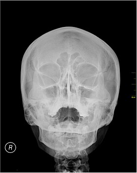
 A Waters' view radiograph showing the paranasal sinuses | |
| Specialty | Radiology |
Waters' view (also known as the occipitomental view or parietoacanthial projection) is a radiographic view of the skull. It is commonly used to get a better view of the maxillary sinuses. An x-ray beam is angled at 45° to the orbitomeatal line. The rays pass from behind the head and are perpendicular to the radiographic plate. Another variation of the waters places the orbitomeatal line at a 37° angle to the image receptor. It is named after the American radiologist Charles Alexander Waters.
Uses
Structures observed
Waters' view can be used to best visualise a number of structures in the skull.
- Maxillary sinuses.
- Frontal sinuses, seen with an oblique view.
- Ethmoidal cells.
- Sphenoid sinus, seen through the open mouth.
- Odontoid process, where if it is just below the mentum, it confirms adequate extension of the head.
The frontal sinus may not show the frontal sinus in detail.[1]
Interpretation of results
| Pathology | Observation |
|---|---|
| None (Normal) |
|
| Maxillary sinusitis[2] | 
|
| Polyp |
|
| Malignancy |

|
Procedure

Typically, the x-ray beam is angled at 45° to the orbitomeatal line.[3] Another variation of the waters places the orbitomeatal line at a 37° angle to the image receptor,[4] or 30°.[5]
History

Waters' view is named after the American radiologist Charles Alexander Waters.[6] It is also known as the occipitomental view.[5]
References
- ^ Freeman, M. Brandon; Harshbarger, Raymond J. (2010). "44 - Fractures of the Frontal Sinus". Plastic Surgery Secrets Plus (2nd ed.). Mosby. pp. 291–294. doi:10.1016/B978-0-323-03470-8.00044-2. ISBN 978-0-323-03470-8.
- ^ a b c d Ruprecht, Axel; Lam, Ernest W. M. (2014). "26 - Paranasal Sinus Diseases". Oral Radiology - Principles and Interpretation (7th ed.). Mosby. pp. 472–491. doi:10.1016/B978-0-323-09633-1.00026-2. ISBN 978-0-323-09633-1.
- ^ Butler, Paul; Mitchell, Adam W. M. (Oct 28, 1999). Applied Radiological Anatomy. p. 97. ISBN 9780521481106.
- ^ Merrill's Atlas of Radiographic Positioning and Procedures. Vol. 2. p. 328.
- ^ a b Archer-Arroyo, Krystal; Mirvis, Stuart E. (2020). "1.2 - Radiological Evaluation of the Craniofacial Skeleton". Facial Trauma Surgery - From Primary Repair to Reconstruction. Elsevier. pp. 16–31. doi:10.1016/B978-0-323-49755-8.00010-4. ISBN 978-0-323-49755-8. S2CID 198282822.
- ^ Chiu, Tor Wo (2019). Stone's plastic surgery facts : a revision guide (4 ed.). Boca Raton, FL: CRC Press. ISBN 978-1-315-18567-5. OCLC 1060523898.
