Condensing osteitis
| Condensing osteitis | |
|---|---|
| Other names | focal sclerosing osteomyelitis |
 | |
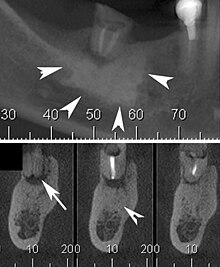
| Cone beam CT scan presenting a diffuse hyperdense lesion in the apex of a mandibular molar (arrowhead, top) adjacent to an inflammatory periapical lesion (arrow, bottom).[1] | |
| Specialty | Dentistry |
Condensing osteitis, also known as focal sclerosing osteomyelitis, is a rare periapical inflammatory condition characterized by the formation of sclerotic bone near the roots of premolars and molars. This condition arises as a response to dental infections, such as periapical pulp inflammation or low-intensity trauma. The lesion typically appears as a radiopacity in the periapical area due to the sclerotic reaction. While most commonly associated with non-vital teeth, condensing osteitis can also occur in vital teeth following occlusal trauma. The condition was first described by Dr. Carl Garré in 1893.[2][3]
Signs and symptoms
Patients are typically asymptomatic, and the condition is most often detected incidentally during routine radiographic examinations.[3] In rare symptomatic cases, patients may report mild pain.[4]
Causes
The cause of condensing osteitis is not clear, but it is thought to happen due to irritation or inflammation that increases osteoblastic activity.[4]
Chronic pulpitis is one of the main factors. This is when bacteria enters the pulp of the tooth, often from untreated cavities. This leads to ongoing mild inflammation that causes bone growth near the root of the tooth.[5] Another main factor is periapical infections. These infections start at the tip of the tooth root, often from dead pulp tissue or occlusal trauma.[6]
Pathophysiology
Condensing osteitis happens when the bone around the tooth reacts to long-term inflammation. This involves excessive bone growth, leading to the formation of sclerotic bone in the jaw.
Ongoing tooth infections, like pulpitis, release chemicals that attract immune cells and activate osteoblasts. Osteoblasts create extra bone in response to inflammation, making the area look more sclerotic. Unlike typical infections, condensing osteitis doesn't destroy bone or produce pus it just adds more bone to the affected area.[4][6]
Diagnosis
Diagnosis typically involves a clinical examination by a dentist or endodontist, complemented by imaging studies such as cone-beam computed tomography. Radiographically, condensing osteitis presents as a localized radiopaque lesion at the root apex of the affected tooth.[7]

Treatment
Condensing osteitis is usually benign and asymptomatic. Treatment depends on the underlying cause and severity. Endodontic treatment is the primary intervention in cases of pulpal infection. Other treatment options include Antibiotics that are prescribed for associated bacterial infections and tooth Extraction: Reserved for cases where the tooth is irreversibly damaged due to pulpitis.[8]
Prognosis
The prognosis is excellent. Following root canal therapy, the sclerotic bone often remodels, restoring normal bone structure over time.[9][10]
Epidemiology
This affects 4-7% of the human population. It mainly occurs during early adolescence/young adults but can occur at any age and in any gender.[11]
Research direction
Recent research has focused on the prevalence and management of condensing osteitis and thus has pointed out the need for further investigation into its pathophysiology and treatment. For instance, studies on the frequency and distribution of mandibular condensing osteitis in specific populations, such as the Taiwanese population, have provided useful epidemiological data. These studies indicate that prevalence may be related to cultural, dietary, or even genetic factors, thus constituting a background for conducting research in different population groups.[11]
Case reports, such as the documentation of a female patient aged 35 years with condensing osteitis, further support that there is variability in clinical presentation and outcome. These cases serve to further the understanding not only of the condition itself but also the deficits in both diagnostic and therapeutic approaches, underlining the need for individual treatment strategies, as well as follow-up over a long period of time regarding changes in bone after intervention.[12]
Future studies may be directed at the molecular mechanisms of osteoblastic activity and bone sclerotization in response to chronic inflammation. Identifying specific inflammatory or genetic markers may lead to earlier diagnosis and targeted therapies. Clinical practices may also be greatly improved by investigating the place of advanced imaging technologies, such as cone-beam computed tomography, in enhancing diagnostic accuracy and monitoring treatment outcomes.
Condensing osteitis will be more clearly understood when epidemiological data is combined with clinical case studies and molecular research to enhance patient care and outcomes by both dental and medical specialties.
References
- ^ Silva, Brunno Santos Freitas; Bueno, Mike Reis; Yamamoto-Silva, Fernanda P.; Gomez, Ricardo Santiago; Peters, Ove Andreas; Estrela, Carlos (2017-07-03). "Differential diagnosis and clinical management of periapical radiopaque/hyperdense jaw lesions". Brazilian Oral Research. 31: e52. doi:10.1590/1807-3107BOR-2017.vol31.0052. PMID 28678971.
- ^ Gumber, Parvind; Sharma, Asmita; Sharma, Kanchan; Gupta, Sonal; Bhardwaj, Bindu; Kant Jakhar, Kamal (December 10, 2024). "Garre's Sclerosing Osteomyelitis – A Case Report" (PDF). [[1]]. Retrieved December 10, 2024.
- ^ a b Goupil, Michael T.; Banki, Mohammad; Ferneini, Elie M. (2016-01-01), Hupp, James R.; Ferneini, Elie M. (eds.), "13 - Osteomyelitis and Osteonecrosis of the Jaws", Head, Neck, and Orofacial Infections, St. Louis: Elsevier, pp. 222–231, doi:10.1016/b978-0-323-28945-0.00013-2, ISBN 978-0-323-28945-0, retrieved 2024-11-06
- ^ a b c Altun, Oğuzhan; Dedeoğlu, Numan; Umar, Esma; Yolcu, Ümit; Acar, Ahmet Hüseyin (2014-01-23). "Condensing Osteitis Lesions in Eastern Anatolian Turkish Population". Oral Surgery, Oral Medicine, Oral Radiology. 2 (2): 17–20. doi:10.12691/oral-2-2-3. ISSN 2333-1135.
- ^ Rechenberg, Dan-Krister; Galicia, Johnah C.; Peters, Ove A. (2016-11-29). Kerkis, Irina (ed.). "Biological Markers for Pulpal Inflammation: A Systematic Review". PLoS One. 11 (11): e0167289. doi:10.1371/journal.pone.0167289. ISSN 1932-6203. PMC 5127562. PMID 27898727.
- ^ a b Neville, Brad W.; Damm, Douglas D.; Allen, Carl M.; Chi, Angela C. (2019-01-01), Neville, Brad W.; Damm, Douglas D.; Allen, Carl M.; Chi, Angela C. (eds.), "3 - Pulp and Periapical Disease", Color Atlas of Oral and Maxillofacial Diseases, Philadelphia: Elsevier, pp. 79–92, doi:10.1016/b978-0-323-55225-7.00003-8, ISBN 978-0-323-55225-7, retrieved 2024-11-06
- ^ Silva, Brunno Santos Freitas; Bueno, Mike Reis; Yamamoto-Silva, Fernanda P.; Gomez, Ricardo Santiago; Peters, Ove Andreas; Estrela, Carlos (2017-07-03). "Differential diagnosis and clinical management of periapical radiopaque/hyperdense jaw lesions". Brazilian Oral Research. 31: e52. doi:10.1590/1807-3107BOR-2017.vol31.0052. ISSN 1807-3107. PMID 28678971.
- ^ "Condensing osteitis". www.pathologyoutlines.com. Retrieved 2024-11-06.
- ^ Eliasson, Sören; Halvarsson, Curt; Ljungheimer, Claes (1984-02-01). "Periapical condensing osteitis and endodontic treatment". Oral Surgery, Oral Medicine, Oral Pathology. 57 (2): 195–199. doi:10.1016/0030-4220(84)90211-1. ISSN 0030-4220.
- ^ Diana, Rasda; Widyastuti, Andina; Santosa, Pribadi (2021-01-16). "One-Visit Apexification in Management of Gutta Percha Extrusion and Condensing Osteitis: 4 Years Follow-Up". Atlantis Press. Atlantis Press: 161–166. doi:10.2991/ahsr.k.210115.034. ISBN 978-94-6239-315-8.
- ^ a b Yeh, Hsiao-Wen; Chen, Ching-Yang; Chen, Pei-Hsuan; Chiang, Meng-Ta; Chiu, Kuo-Chou; Chung, Ming-Pang; Li, Tsung-I; Su, Chi-Chun; Shieh, Yi-Shing (2015-09-01). "Frequency and distribution of mandibular condensing osteitis lesions in a Taiwanese population". Journal of Dental Sciences. 10 (3): 291–295. doi:10.1016/j.jds.2014.10.002. ISSN 1991-7902.
- ^ Tsvetanov, Tsvetan. "Mandibular Idiopathic Osteosclerosis or Condensing Osteitis. A Case Report" (PDF). researchgate.
External links
Category:Inflammations Category:Osteitis Category:Pathology of the maxilla and mandible
