Neuromelanin

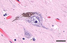
Neuromelanin (NM) is a dark pigment found in the brain which is structurally related to melanin. It is a polymer of 5,6-dihydroxyindole monomers.[1] Neuromelanin is found in large quantities in catecholaminergic cells of the substantia nigra pars compacta and locus coeruleus, giving a dark color to the structures.[2]
Physical properties and structure

Neuromelanin gives specific brain sections, such as the substantia nigra or the locus coeruleus, distinct color. It is a type of melanin and similar to other forms of peripheral melanin. It is insoluble in organic compounds, and can be labeled by silver staining. It is called neuromelanin because of its function and the color change that appears in tissues containing it. It contains black/brown pigmented granules. Neuromelanin is found to accumulate during aging, noticeably after the first 2–3 years of life. It is believed to protect neurons in the substantia nigra from iron-induced oxidative stress. It is considered a true melanin due to its stable free radical structure and it avidly chelates metals.[3]
Synthetic pathways
Neuromelanin is directly biosynthesized from L-DOPA, precursor to dopamine, by tyrosine hydroxylase (TH) and aromatic acid decarboxylase (AADC). Alternatively, synaptic vesicles and endosomes accumulate cytosolic dopamine (via vesicular monoamine transporter 2 (VMAT2) and transport it to mitochondria where it is metabolized by monoamine oxidase. Excess dopamine and DOPA molecules are oxidized by iron catalysis into dopaquinones and semiquinones which are then phagocytosed and are stored as neuromelanin.[4]
Neuromelanin biosynthesis is driven by excess cytosolic catecholamines not accumulated by synaptic vesicles.[5]
Function
Neuromelanin is found in higher concentrations in humans than in other primates.[2] Neuromelanin concentration increases with age, suggesting a role in neuroprotection (neuromelanin can chelate metals and xenobiotics[6]) or senescence.[citation needed]
Role in disease
Neuromelanin-containing neurons in the substantia nigra degenerate during Parkinson's disease.[citation needed] Motor symptoms of Parkinson's disease are caused by cell death in the substantia nigra, which may be partly due to oxidative stress.[citation needed] This oxidation may be relieved by neuromelanin.[citation needed] Patients with Parkinson's disease had 50% the amount of neuromelanin in the substantia nigra as compared to similar patients of their same age, but without Parkinson's.[citation needed] The death of neuromelanin-containing neurons in the substantia nigra, pars compacta, and locus coeruleus have been linked to Parkinson's disease and also have been visualized in vivo with neuromelanin imaging.[7]
Neuromelanin has been shown to bind neurotoxic and toxic metals that could promote neurodegeneration.[5]
History
Dark pigments in the substantia nigra were first described in 1838 by Purkyně,[8] and the term neuromelanin was proposed in 1957 by Lillie,[9] though it has been thought to serve no function until recently. It is now believed to play a vital role in preventing cell death in certain parts of the brain. It has been linked to Parkinson's disease and because of this possible connection, neuromelanin has been heavily researched in the last decade.[10]
References
- ^ Charkoudian LK, Franz KJ (2006). "Fe(III)-coordination properties of neuromelanin components: 5,6-dihydroxyindole and 5,6-dihydroxyindole-2-carboxylic acid". Inorganic Chemistry. 45 (9): 3657–64. doi:10.1021/ic060014r. PMID 16634598.
- ^ a b Fedorow, H; Tribl, F; Halliday, G; Gerlach, M; Riederer, P; Double, K. L. (2005). "Neuromelanin in human dopamine neurons: Comparison with peripheral melanins and relevance to Parkinson's disease". Progress in Neurobiology. 75 (2): 109–24. doi:10.1016/j.pneurobio.2005.02.001. PMID 15784302. S2CID 503902.
- ^ "IBA in life sciences". Archived from the original on 2013-12-03. Retrieved 2013-11-30.
- ^ Rabey, J.M.; Hefti, F. (1990). "Neuromelanin synthesis in rat and human substantia nigra". Journal of Neural Transmission. Parkinson's Disease and Dementia Section. 2 (1): 1–14. doi:10.1007/BF02251241. PMID 2357268. S2CID 6769760.
- ^ a b Stepień, K; Dzierzega-Lecznar, A; Tam, I (2007). "The role of neuromelanin in Parkinson's disease--new concepts". Wiadomosci Lekarskie. 60 (11–12): 563–9. PMID 18540183.
- ^ Tribl, F; Asan, E; Arzberger, T; Tatschner, T; Langenfeld, E; Meyer, H. E.; Bringmann, G; Riederer, P; Gerlach, M; Marcus, K (2009). "Identification of L-ferritin in neuromelanin granules of the human substantia nigra: A targeted proteomics approach". Molecular & Cellular Proteomics. 8 (8): 1832–8. doi:10.1074/mcp.M900006-MCP200. PMC 2722774. PMID 19318681.
- ^ Sasaki M, Shibata E, Tohyama K, Takahashi J, Otsuka K, Tsuchiya K, Takahashi S, Ehara S, Terayama Y, Sakai A (July 2006). "Neuromelanin magnetic resonance imaging of locus ceruleus and substantia nigra in Parkinson's disease". NeuroReport. 17 (11): 1215–8. doi:10.1097/01.wnr.0000227984.84927.a7. PMID 16837857. S2CID 24597825.
- ^ Usunoff, K. G.; Itzev, D. E.; Ovtscharoff, W. A.; Marani, E (2002). "Neuromelanin in the human brain: A review and atlas of pigmented cells in the substantia nigra". Archives of Physiology and Biochemistry. 110 (4): 257–369. doi:10.1076/apab.110.4.257.11827. PMID 12516659. S2CID 2735201.
- ^ Lillie, R. D. (1957). "Metal reduction reactions of the melanins: Histochemical studies". Journal of Histochemistry and Cytochemistry. 5 (4): 325–33. doi:10.1177/5.4.325. PMID 13463306.
- ^ Zecca, L; Tampellini, D; Gerlach, M; Riederer, P; Fariello, R. G.; Sulzer, D (2001). "Substantia nigra neuromelanin: Structure, synthesis, and molecular behaviour". Molecular Pathology. 54 (6): 414–8. PMC 1187132. PMID 11724917.
